Seeing the Unseen: How Brain Scans Help Diagnose Alzheimer’s Disease
Share IT

Launch Your Dream Website with Us!
Click Here to Get in touch with Us.
Categories
Brain Scans for Alzheimer’s Diagnosis
Uncovering the Brain: Alzheimer’s Disease Neuroimaging Methods Identification
The most prevalent type of dementia, Alzheimer’s disease (AD), poses a serious challenge to medical personnel as well as individuals. Timely management and enhanced patient outcomes are contingent upon an early and precise diagnosis. Although cognitive tests are still the mainstay of AD diagnosis, neuroimaging methods have become potent diagnostic instruments.
Brain Scans for Alzheimer’s Diagnosis
This blog post explores the field of neuroimaging for the diagnosis of AD, looking at different methods, uses, benefits, and drawbacks.
Table of Contents
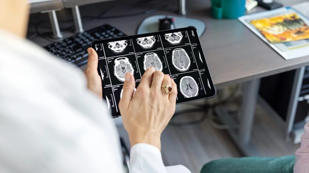
Methods for Revealing the Brain’s Topography
Brain Scans for Alzheimer’s Diagnosis
1. Structural Imaging:
When it comes to displaying the structure of the brain, magnetic resonance imaging (MRI) is the gold standard. Particularly in the hippocampus, an essential organ for memory, it can identify atrophy, or shrinking, in particular brain regions, which is a characteristic of AD. Diffusion-weighted MRI is one of the advanced MRI techniques that can evaluate white matter integrity and possibly identify early indicators of neurodegeneration.
Brain Scans for Alzheimer’s Diagnosis
Computed Tomography (CT) scans are widely available and can rule out alternative causes of dementia with comparable symptoms, such as strokes or tumours. However, they are less sensitive than MRIs in identifying subtle changes.
Brain Scans for Alzheimer’s Diagnosis
- Imaging Functions:
Positron Emission Tomography (FDG-PET): This imaging technique assesses the metabolism of glucose in the brain. Reduced glucose uptake is seen in AD patients, which is indicative of diminished neuronal activity. FDG-PET is a useful method for identifying AD from other dementias, however it is not able to identify the underlying pathology.
- Imaging Molecular Systems:
- Amyloid PET: A defining feature of AD are amyloid plaques, which are protein deposits in the brain. Tracers that bind to these plaques are used in amyloid PET scans to visualise their extent. This does not always indicate the severity of dementia, but it does help validate the existence of AD pathology.
- Tau PET: By employing particular tracers, tau tangles—an additional protein anomaly associated with AD—can also be seen on PET scans. Although tau PET imaging is still in its early stages of research, it has the potential to distinguish AD from other neurodegenerative illnesses.
- Benefits of Neuroimaging for AD Diagnosis: Increased Accuracy: Neuroimaging can be used to objectively demonstrate changes in the brain, which can help with diagnosis, especially in the early stages when symptoms may be mild.
- Differential Diagnosis: Neuroimaging can assist in differentiating Alzheimer’s disease (AD) from other dementias that share symptoms by highlighting certain patterns of brain abnormalities.
- Tracking the Progression of the Illness: Regular neuroimaging scans can track the advancement of an illness over time, enabling modifications to treatment regimens.
- Research and Development: In order to comprehend the disease process and create new treatments, neuroimaging is essential to AD research.
- Cost and Accessibility Limitations: Neuroimaging methods can be costly, and not all healthcare facilities have easy access to them.
- Radiation Exposure: Certain procedures, such as PET scans, expose patients to low doses of radiation, which some patients may find concerning.
- Interpretation Difficulties: Determining the meaning of neuroimaging results can be difficult and need specialised knowledge. Furthermore, some modifications might not be exclusive to AD.
Neuroimaging’s Prospects in AD Diagnosis
Brain Scans for Alzheimer’s Diagnosis
Neuroimaging for AD diagnosis is ripe for improvement as technology develops further:
- Artificial Intelligence and Machine Learning: Machine learning algorithms have the ability to analyse neuroimaging data more accurately and efficiently, which could result in earlier and more accurate diagnoses.
- Creation of New Tracers: The sensitivity and specificity of neuroimaging methods may be enhanced by the creation of more targeted tracers for various pathological indicators of AD.
- Multimodal Imaging: By combining information from several neuroimaging modalities, a more complete picture of the illness may be obtained, enabling a more individualised approach to diagnosis and therapy.
In Summary:
Brain Scans for Alzheimer’s Diagnosis
neuroimaging has emerged as a crucial diagnostic tool for Alzheimer’s disease. Even with their limits, current developments hold great promise for the future. By utilising these developments, we can get closer to a future in which AD can be successfully identified and treated, enhancing the quality of life for both patients and their families.

Launch Your Dream Website with Us!
Click Here to Get in touch with Us.












Recent Comments