COPD Imaging Tests Explained
Peering Inside: CT Scan and Chest X-Ray Results in COPD
Breathing difficulties are a hallmark of chronic obstructive pulmonary disease (COPD), a lung disorder that progresses over time. Imaging is essential for both COPD diagnosis and treatment. This blog post discusses the limits of chest X-rays and CT scans in diagnosing COPD and how they might be used to find anomalies related to the disease.
Table of Contents

A First Look at a Chest X-Ray
COPD Imaging Tests Explained
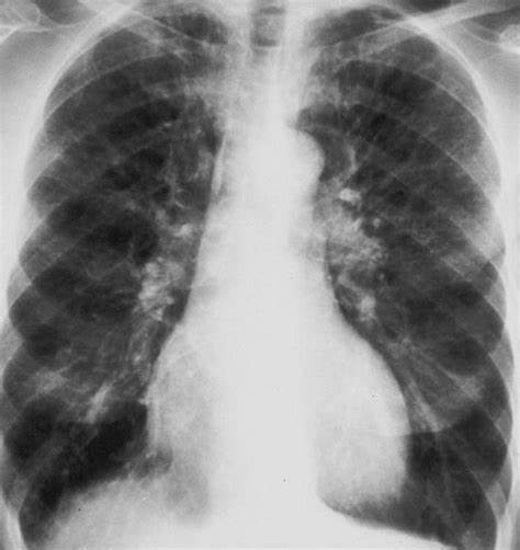
Usually, the first imaging test performed to assess potential lung issues is a chest X-ray. Chest X-rays can show some anomalies suggestive of COPD, albeit they are not conclusive in the diagnosis of the condition. These anomalies include:
- Hyperinflation: Air trapping may cause the lungs to seem larger than normal.
- Flattened Diaphragm: When the lungs expand too much, the diaphragm, which is a muscle that separates the chest from the abdomen, gets pushed upward and appears flattened.
- Increased Bronchovascular Markings: Inflammation can make blood vessels and airways stand out more.
- Bullae: When lung tissue is destroyed, these big air pockets can develop in the lungs. Large bullae may be visible on X-rays, but tiny ones may not be picked up.
Restrictions on the Use of Chest X-Rays to Diagnose COPD:
- Non-specific findings: Heart failure or asthma are two more lung disorders that might induce changes on chest X-rays.
- Limited sensitivity: X-rays taken in the early stages of COPD may not reveal any appreciable abnormalities.
- Emphysema assessment difficulty: Emphysema is a prominent component of COPD that chest X-rays may not be able to definitively identify.
CT Scan: A Closer Exam
COPD Imaging Tests Explained
Comprehensive cross-sectional images of the lungs and adjacent tissues can be obtained with a chest CT scan. Although they are not usually utilized for the first diagnosis, CT scans provide additional details regarding the structure of the lungs and can be useful for:
- Verifying Emphysema: By identifying regions of low lung density, CT scans can detect and measure emphysema.
- Assessing the Severity of the Disease: CT scans can help determine the severity of a disease by giving a more detailed image of lung damage and airway obstruction.
- Distinguishing Other Conditions: CT scans can assist in excluding other lung disorders that may resemble the symptoms of COPD.
- therapy Planning: In situations of severe COPD, lung volume reduction surgery is a potential therapy option that can be planned with the assistance of CT scans.
CT Scan Restrictions for COPD Diagnosis:
- Radiation exposure: Because CT scans need ionizing radiation exposure, they are not recommended for routine screening.
- High cost: Compared to chest X-rays, CT scans are usually more costly.
- Overdiagnosis: Small abnormalities that may not be clinically important may be found on CT scans.
The Value of an All-encompassing Strategy
COPD Imaging Tests Explained
In the diagnosis and treatment of COPD, chest X-rays and CT scans are useful tools, but they should be interpreted in combination with additional information, such as:
- Symptoms: Common signs of COPD include tightness in the chest, coughing, and dyspnea.
- Medical history: Two important risk factors for COPD are a history of smoking and exposure to lung irritants.
- Physical examination: A medical practitioner can evaluate breathing patterns and lung sounds.
- Spirometry: A test of pulmonary function that measures airflow limitation—a defining feature of COPD—is available.
COPD Imaging Tests Explained
Healthcare professionals can accurately diagnose COPD and create a suitable treatment strategy by integrating imaging results with other clinical data.
COPD Living: Beyond Imaging
COPD Imaging Tests Explained
Although imaging is useful in managing COPD, the following should be prioritized:
- Symptom Management: To assist control symptoms and enhance quality of life, doctors may prescribe medication, oxygen therapy, or pulmonary rehabilitation.
- Cessation of Smoking: The single most critical step in reducing the course of COPD is quitting smoking.
- vaccines: To avoid respiratory infections that exacerbate COPD, routine vaccines against the flu and pneumonia are essential.
COPD Imaging Tests Explained
For those with COPD, early diagnosis, thorough treatment, and proactive lifestyle changes can greatly enhance quality of life.


GIPHY App Key not set. Please check settings