Heart Imaging Tests
Seeing Clearly Within: Imaging Methods for Diagnosing Heart Disease
The best treatment is due to the heart, the unceasing engine that powers our existence. To keep your heart healthy, it’s important to find any underlying problems early. Thanks to developments in medical technology, we now have access to a variety of imaging modalities that allow us to see clearly inside the chest and identify any potential issues with the heart or surrounding structures. This blog post examines the four main imaging modalities—chest X-ray, CT scan, MRI, and nuclear imaging—that are used to diagnose cardiac disease.
Table of Contents

- A brief overview of the chest using a chest X-ray
A chest X-ray gives a basic image of the chest cavity and is a simple, painless, and accessible imaging procedure. Although less comprehensive than other methods, it can provide insightful information for the diagnosis of heart disease by disclosing:Heart Imaging Tests
- Size and shape of the heart: An oversized or deformed heart may be a sign of underlying cardiac diseases, such as heart failure.
- Lung congestion: An X-ray of the chest can reveal fluid accumulation in the lungs, which may indicate heart failure.
- Aortic dissection: Sometimes seen on a chest X-ray, this potentially fatal ailment is caused by a tear in the aorta, the main artery leaving the heart.
Heart Imaging Tests
Chest X-rays do have certain limits, though. The coronary arteries, which provide blood to the heart muscle, are not visible to them directly. Furthermore, they might not always offer sufficient information to make a firm diagnosis of a particular cardiac problem.
- CT Scan: A Comprehensive Exam of the Heart and Blood Vessels
A non-invasive imaging method called computed tomography (CT) scan makes use of X-rays to produce finely detailed cross-sectional images of the chest. A CT scan offers a more thorough image than a conventional X-ray, revealing:Heart Imaging Tests
- Heart anatomy: It is possible to see the dimensions, form, and chambers of the heart in great detail.
- Coronary arteries: To check for blockages in the coronary arteries, a CT scan with contrast dye, a specific dye injected into the bloodstream, may be utilized.
- Aortic health: Aneurysms (bulges), which are anomalies of the major artery that carries blood out from the heart, can be checked for.
Heart Imaging Tests
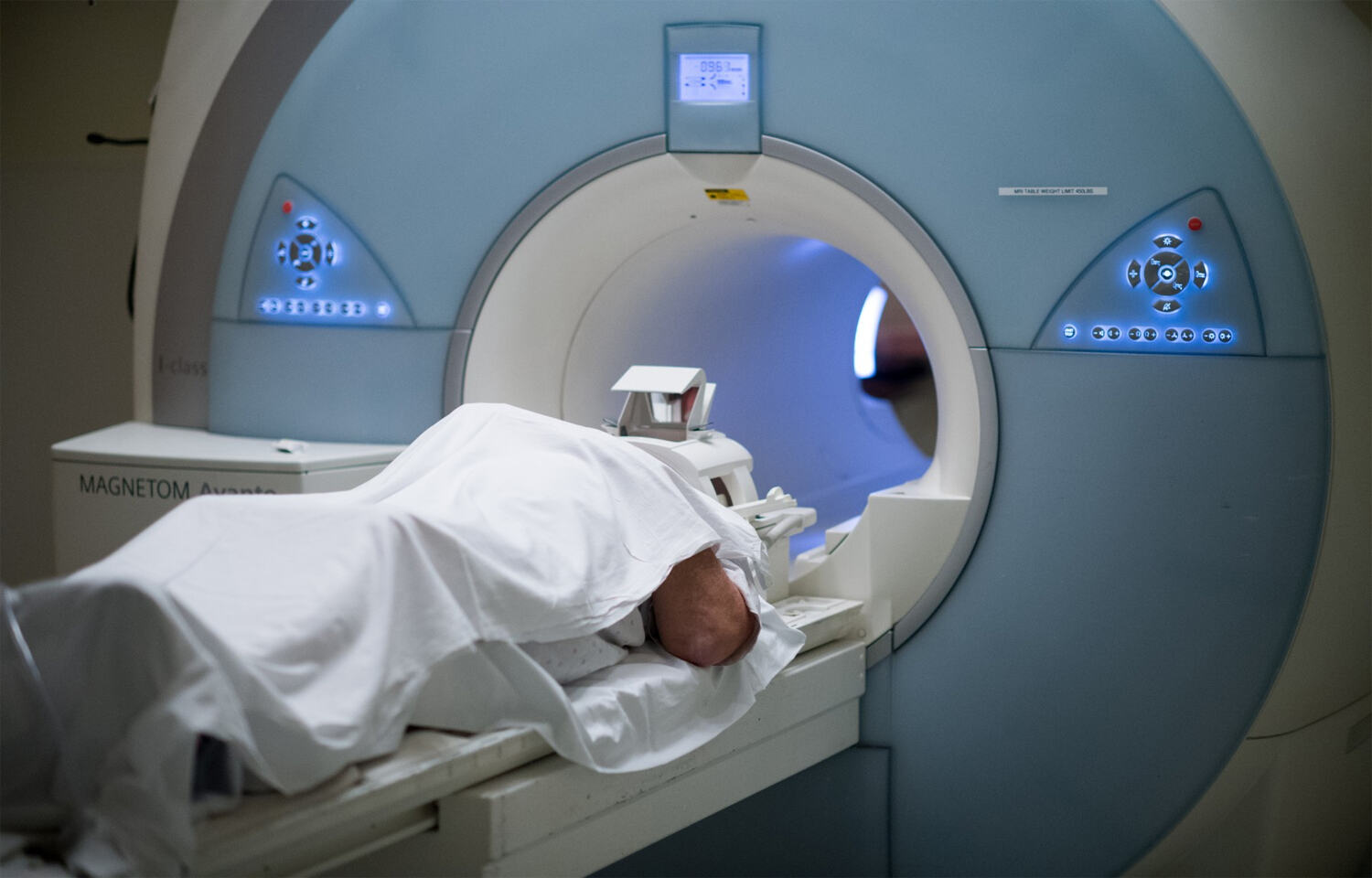
- MRI: Revealing the Structure and Function of the Heart
Strong magnetic fields and radio waves are used in magnetic resonance imaging (MRI), a potent imaging method that produces finely detailed images of the heart and surrounding tissues. MRIs do not employ radiation, in contrast to CT scans. An MRI examination can show:
- Heart anatomy: An MRI, like a CT scan, offers fine-grained pictures of the heart’s composition, including its chambers, valves, and surrounding tissues.
- Heart function: An MRI can evaluate the heart’s pumping efficiency by measuring how well the heart muscle pumps and relaxes with each beat.
- Abnormalities of the heart wall: MRI can identify conditions such as inflammation or scarring of the heart muscle.
Heart Imaging Tests
MRI scans are more costly and time-consuming than other methods, but they are also very insightful. Moreover, claustrophobia sufferers and those with specific medical implants should not have MRIs.
- Nuclear Imaging: Evaluation of Blood Movement and Purpose
A sophisticated method called nuclear imaging involves injecting radioactive tracers into the bloodstream. By accumulating in particular tissues, these tracers help medical professionals evaluate blood flow and function. When diagnosing cardiac illness, nuclear imaging often uses two types of images:Heart Imaging Tests
- Myocardial perfusion imaging (MPI): A technique called myocardial perfusion imaging (MPI) evaluates blood flow to the heart muscle both at rest and under stress (from exercise or medicine). A blocked artery may be indicated by decreased blood flow.
- Positron emission tomography (PET) scan for the heart: Like MPI, a PET scan evaluates heart function and blood flow using radioactive tracers. Additionally, following a cardiac attack, it can offer details regarding the viability of the heart muscle (living tissue).
Heart Imaging Tests
Tests using nuclear imaging provide important details regarding the function and flow of blood in the heart muscle. Nonetheless, they entail minimal radiation exposure and might not be easily accessible in every medical facility.
Selecting the Appropriate Imaging Method
Heart Imaging Tests
The suspected heart problem, your medical history, and the accessibility of various technologies all play a role in the imaging approach chosen for the diagnosis of heart illness. After taking these things into account, your doctor will suggest the test that would best meet your needs.
Recall that effective management of cardiac disease depends on early detection. You should speak with your doctor about potential risk factors and suitable diagnostic testing if you have any concerns about the health of your heart.


Recent Comments